Обследования на рак эндометрия (матки)
Обследования на рак эндометрия (рак матки ) рекомендуется только женщинам из группы повышенного риска.
Повышенный риск не означает, что вы обязательно заболеете раком эндометрия. Но вам необходимо начать проходить регулярные обследования с целью профилактики и раннего выявления опухолевых заболеваний. При раннем обнаружении рака шансы на успешное лечение значительно высоки.
Факторы риска:
Женщины, к которым относится хоть один из указанных факторов риска, должны регулярно проходить обследования на рак эндометрия:
Наряду с регулярными обследованиями необходимо наблюдать за своим состоянием. В случае таких симптомов, как нерегулярные кровотечения или выделения, следует незамедлительно сообщить о них врачу.
Для женщин, перенесших рак эндометрия, необходим индивидуальный план обследования на рецидив, составленный лечащим врачом.
В ННОЦ вы можете пройти качественную диагностику, выполнить «пересмотр стеклопрепаратов» для получения «второго мнения» о типе клеток опухоли, а при необходимости, безотлагательно начать и получить квалифицированное лечение.
Граждане Казахстана имеют право свободного выбора лечащего врача и медицинской организации согласно подпункту 3, пункта 1, Статьи 77 Кодекса Республики Казахстан «О здоровье и системе здравоохранения».
Кроме того, пациент имеет право свободного выбора организации здравоохранения при плановой госпитализации, что закреплено приказом Министра здравоохранения и социального развития Республики Казахстан от 29 сентября 2015 года № 761 «Об утверждении Правил оказания стационарной помощи».
Обследования на рак яичников
В настоящее время обследования на рак яичников рекомендуется только женщинам из группы повышенного или высокого риска.
При этом, повышенный риск не означает, что вы обязательно заболеете раком яичников. Но вам может потребоваться начать регулярные осмотры и обследования. Поэтому, если вы заболели раком, ваш врач обнаружит его на самой ранней стадии. При раннем обнаружении шансы на успешное излечение болезни велики.
Наряду с регулярными обследованиями наблюдайте за своим состоянием. Если вы заметили некоторые симптомы, такие как дискомфорт в животе или боль, увеличение живота в объеме, незамедлительно обратитесь к врачу, который направит на необходимые обследования.
Вам следует пройти обследование на рак яичников, если у вас есть один из нижеследующих факторов риска:
Если вы попадаете в какую-либо из этих групп риска, вам следует проходить:
Если у вас подтвердился рак, согласуйте с вашим лечащим врачом ваш план лечения. Если же у вас был рак яичников и вы прошли лечение обязательно согласуйте ваш план обследований на рецидив.
В ННОЦ вы можете пройти качественную диагностику, получить «второе мнение» о типе клеток опухоли при поставленном диагнозе, а при необходимости, безотлагательно начать и получить квалифицированное лечение.
Граждане Казахстана имеют право свободного выбора лечащего врача и медицинской организации согласно подпункту 3, пункта 1, Статьи 77 Кодекса Республики Казахстан «О здоровье и системе здравоохранения».
Кроме того, пациент имеет право свободного выбора организации здравоохранения при плановой госпитализации, что закреплено приказом Министра здравоохранения и социального развития Республики Казахстан от 29 сентября 2015 года № 761 «Об утверждении Правил оказания стационарной помощи».
Ультрафиолетовое излучение — вид электромагнитного излучения. Главным источником ультрафиолета являются солнечные лучи, а также искусственные источники УФ-излучения, например, в соляриях.
УФ-излучение является источником радиации – менее сильным, чем, например, рентгеновские лучи, но более сильным чем радиоволны. Это свойство придает УФ лучам способность забирать электрон из атома или молекулы, т. е. ионизировать (поэтому радиацию называют ионизирующей). Ионизирующая радиация способна повреждать ДНК клеток, что может вызывать рак кожи. Одним из самых важных и простых способов снизить риск рака кожи - это защитить ее от УФ-излучения. Солярии также излучают УФ-излучение и вызывают такое же повреждение кожи, как если бы вы загорали под солнцем.
Виды рака кожи, которые способно вызвать ультрафиолетовое облучение:
Для защиты кожи от вредного воздействия ультрафиолетовых лучей врачи рекомендуют:
Признаки опухолевых заболеваний кожи:
Существует общепризнанный алгоритм оценки пигментных образований ABCDE, который может помочь определению «подозрительных». Он состоит из первых букв оцениваемых признаков:
Если у вас имеются вышеперечисленные симптомы или вы заметили подозрительные образования на коже, незамедлительно обратитесь к врачу.
Myoma is the most common type of tumor of the female reproductive organs and is the most common type of tumor of the female reproductive organs, which occurs in 20-40% of women of reproductive age.
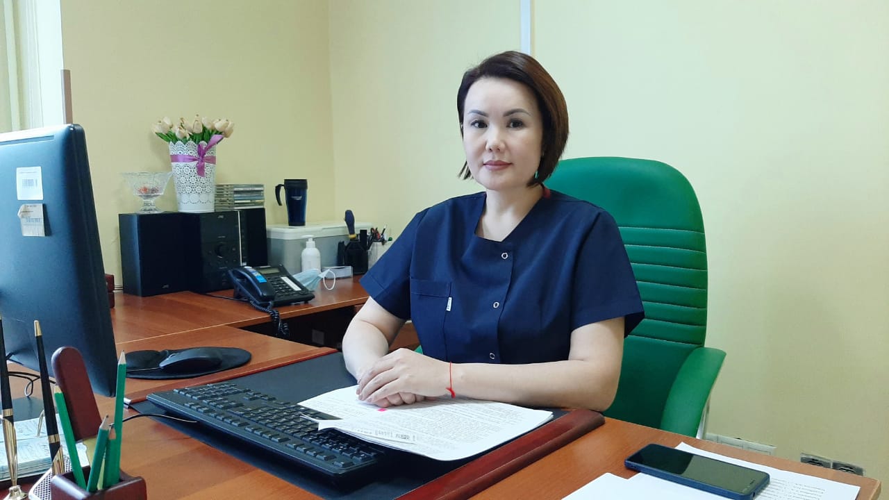
![]() Kulkeva Gulnar Utepbergenova
Kulkeva Gulnar Utepbergenova
Chairman of the Board of the National oncology research center.
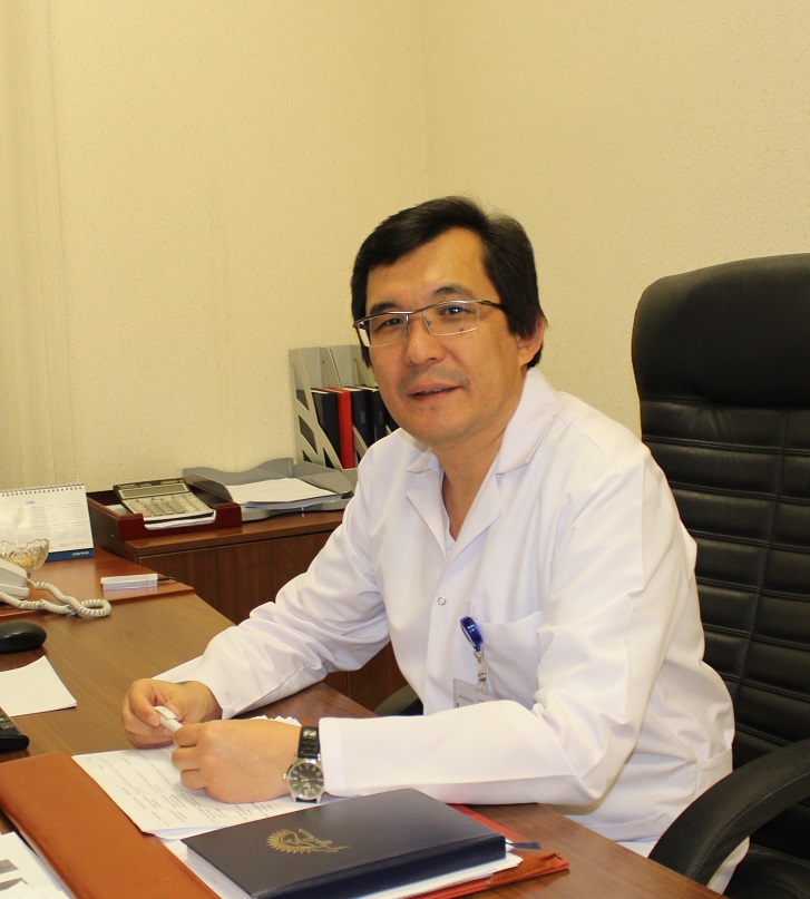 Adilbek Kairbekovich Mukazhanov
Deputy Chairman Of the Board for medical activities.
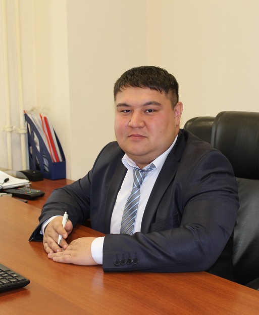 Satybaev Kanat Syzdykovich
Deputy Chairman of the Management Board for administrative, legal, financial and economic activities
What is screening?
Screening is a complex of measures, including a preventive medical examination and additional examination methods, carried out in order to detect a tumor at an early stage, when there are no symptoms of the disease yet.
A tumor diagnosed at an early stage can be completely healed.
At later stages, when there are already symptoms of the disease and there is a possibility of the spread of the tumor, the treatment of the disease becomes more difficult. Therefore, early detection and treatment of a tumor, before its spread, is the most effective way to reduce mortality from malignant neoplasms.
It often happens that the tumor cannot always be detected in the early stages, since the early course of the disease is asymptomatic, the patient does not feel changes in his state of health and does not go to doctors for examination.
It is important to note that being screened does not mean cancer is suspected.
In Kazakhstan, there are screening programs for specific diseases and for certain age groups, which are completely free for the population.
Screening types:
• Breast cancer - women between 40 and 70 years old.
• Cervical cancer - women between 30 and 70 years old.
• Colorectal screening - men and women from 50 to 70 years old.
Screening can be done at the clinic at the place of attachment free of charge.
It is important to understand that prevention, early diagnosis, screening and treatment are key components of successful cancer control. Early detection of cancer saves lives, reduces treatment costs and preserves the quality of life of those who are ill.
Colonoscopy - what is it?
It is well known that such a complex procedure as a colonoscopy is necessary to diagnose pathologies of the colon and rectum. The procedure consists in inserting a probe (colonoscope) into the anus. The probe attaches to a flexible tube and contains a tiny video camera that allows the doctor to examine the mucosal surface throughout the colon. The length of the device is up to 1.45 m, which allows examination of the blind, sigmoid, colon and rectum. Colonoscopy does not involve the small intestine. If necessary, colonoscopy can also remove polyps or other types of growths or take tissue samples for analysis (biopsy).
Why is colonoscopy needed?
The patient is indicated for a colonoscopy for chronic abdominal pain, bleeding from the anus, chronic constipation, diarrhea and other bowel problems. You can also have a colonoscopy to check for bowel cancer. If the patient is over 50 years old and has no history of colon cancer in the family, the doctor may recommend that the patient undergo an abdominal examination with a colonoscope every 7-10 years. If the patient is prone to the formation of polyps on the walls of the large intestine, colonoscopy should be carried out at regular intervals in order to find and remove the polyps that have arisen during the examination of the walls. This manipulation is performed to reduce the risk of rectal cancer.
Preparing for a colonoscopy.
Colonoscopy is a low-traumatic type of medical procedure; most people experience it under the influence of anesthesia. The research itself takes no more than an hour, and recovery after a couple of hours. Preparation for the study is the most difficult for patients. It is a little consoling that if the procedure is successful, and no pathologies have been identified, then the next one may not be required for 7-10 years.
It is well known that colonoscopy requires a preliminary cleansing of the intestines so that it is empty and as clean as possible (as possible) - otherwise food debris and feces can make it difficult for the doctor to see. As a rule, when a doctor prescribes a diagnostic procedure, he tells the patient exactly how to prepare for it, what to eat, what drugs to use and what reactions of the body should be expected in the preparation process. The method of preparation for colonoscopy is selected for each patient individually.
Make an appointment and more detailed information on bowel preparation by phone 8 (7172) 702895.
What is bronchoscopy?
Bronchoscopy is a modern diagnostic study of the mucous membranes of the trachea and bronchi using a special optical device - a bronchoscope. This is the only method that allows you to directly assess the inner surface of the bronchi, to study their configuration, the relief of the mucous membrane and its vascular pattern, and if a pathologically altered area of the mucous membrane is detected, a biopsy is performed for subsequent morphological analysis. Bronchoscopy is also the most important and effective way of treating patients with chronic inflammatory and purulent lung diseases.
Preparation for research.
The study is carried out strictly on an empty stomach, food intake for 8-10 hours and liquid for 4-6 hours before the start of the procedure is completely excluded. In the evening before the study (until 18:00) - a light dinner. On the day of the study, you should refrain from smoking.
Cancel oral anticoagulants (blood thinners) on the eve of the study, pause subcutaneous administration of heparin 4-6 hours before the procedure. For examination, you must have an outpatient card, the results of a CT scan of the chest or a description of the X-ray of the lungs, a towel (since after the procedure, a short hemoptysis is possible). If you suffer from bronchial asthma, do not forget the inhaler. During the preliminary conversation, inform your doctor about your allergy to medicines (especially if you are allergic to pain medications) and your chronic diseases (bronchial asthma, heart failure).
Additional diagnostic and therapeutic manipulations during bronchoscopy:
Mucosal biopsy / neoplasm
Flushing from the walls of the bronchi
Bronchoalveolar lavage
Sanitation of the tracheobronchial tree
What complications can there be?
As a rule, this study is well tolerated by patients, but sometimes there is a loss or hoarseness of the voice, sore throat, and in the case of a biopsy, hemoptysis may occur. These phenomena are temporary. You should be alerted to prolonged hemoptysis, intense unrelenting chest pain, swelling on the face and around the neck, nausea and vomiting, as well as fever and chills. If these symptoms appear, consult a doctor immediately.


